
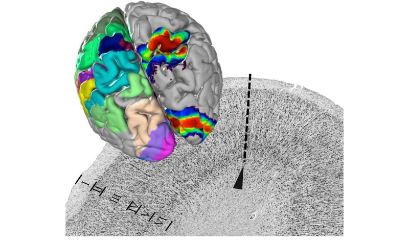
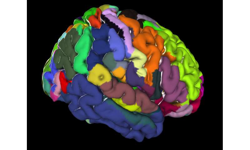
Julich-Brain is the name of the first 3-D-atlas of the human brain that reflects the variability of the brain’s structure with microscopic resolution. The atlas features close to 250 structurally distinct areas, each one based on the analysis of 10 brains. More than 24,000 extremely thin brain sections were digitized, assembled in 3-D and mapped by experts. As part of the new EBRAINS infrastructure of the European Human Brain Project, the atlas serves as an interface to link information about the brain in a spatially precise way. German researchers led by Prof. Katrin Amunts have now presented the new brain atlas in the journal Science.
Under the microscope, it can be seen that the human brain is not uniformly structured, but divided into clearly distinguishable areas. These areas differ in the distribution and density of nerve cells and in function. With the Julich-Brain, researchers led by Katrin Amunts now present the most comprehensive digital map of the brain’s cellular architecture and make it available worldwide via the EBRAINS research infrastructure.
“On the one hand, the digital brain atlas will help to interpret the results of neuroimaging studies, for example, of patients, more accurately,” says Katrin Amunts, director at the German Research Center Juelich and Professor at the University of Duesseldorf. “On the other hand, it is becoming the basis for a kind of ‘Google Earth’ of the brain—because the cellular level is the best interface for linking data about very different facets of the brain.”
A Google Earth of the Brain
In this way, the researchers are making a significant contribution to the Human Brain Project (HBP). “Together with many partners in this project, we are building EBRAINS as a novel, high-tech research infrastructure for the neurosciences,” says Amunts, who is also the scientific research director of the project.
More than a quarter-century of research has gone into the 3-D atlas. Dozens of experts have used image analysis and mathematical algorithms to evaluate the tissue sections over the years and determine the boundaries between brain areas, which together make up a length of almost 2000 meters.
Regions vary in their difference
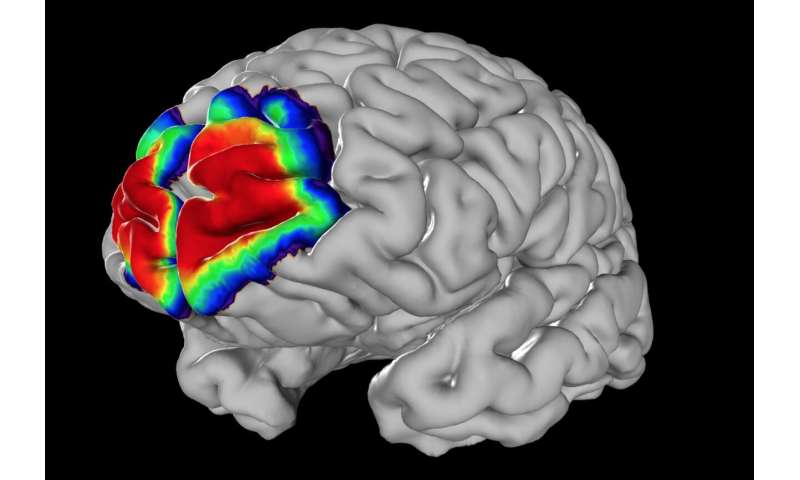
Mapping showed that areas vary between different brains, for example, in terms of size and location. The Julich-Brain therefore displays the position and shape of individual regions as probability maps. The researchers found particularly large differences in the Broca region, which is involved in language. In contrast, the primary visual area appeared much more uniform.
As part of EBRAINS, the Julich Brain Atlas is the starting point for bringing structure and function together. The atlas is already helping to link data on gene expression, connectivity and functional activity to better understand brain functions and the mechanisms of diseases. “EBRAINS also enables us to use the maps for simulations or to apply artificial intelligence to explore the division of labor between brain areas. The huge amounts of data generated from this are processed using the EBRAINS computing platform.” The computational power comes from the new European supercomputing network FENIX, which is formed by five leading centers for high performance computing, including the Julich Supercomputing Center (JSC).
Digital brain science
Source: Read Full Article