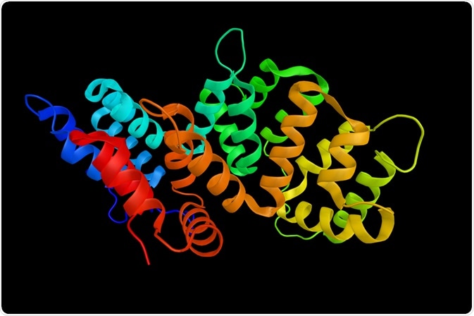
Electron spin resonance spectroscopy (ESR) of spin-labeled molecules is emerging as a potent method to assess the structure and conformation of proteins and nucleic acids.
This article will cover:
- Electron Spin Resonance Spectroscopy (ESR)
- Methods of spin labeling
- Spin-labeling cysteine
- Incorporating spin-labeled amino acids into proteins
- Elucidating structural information from ESR spectra
 ibreakstock | Shutterstock
Electron Spin Resonance Spectroscopy (ESR)
In ESR, a spin label is added to the target site using cysteine substitution. This is followed by modifying the sulfhydryl group with a paramagnetic nitroxide reagent. The ESR then provides information about the mobility of nitroxide side chain, accessibility of the solvent, distance between nitroxide and other paramagnetic centers.
Novel chemistry of probes in terms of incorporation into proteins, labeling, similarity to amino acid chains, and minimal perturbation of the structure and function of proteins have led to advances in monitoring the protein backbone and microenvironment of the probe.
Methods of spin labeling
There are two main methods of modifying the proteins using paramagnetic spin labels. One method involves attaching various nitroxides to the sulfhydryl group of the cysteine residue present in the protein to create a spin label side chain. This method requires that the presence of cysteine residues only at the desired sites, so any extra cysteines in the protein are replaced by serine or alanine.
One of the most frequently used spin label is (1-oxyl-2,2,5,5-tetramethylpyrroline-3 methyl)methanethiosulfonate (MTS) because of its specificity and small molecular volume. Also, the peperidine-oxyl moiety and protein backbone link are flexible, allowing the folding of proteins into native conformations.
However, with this spin label, a distance distribution is obtained, rather than a defined distance. Thus, a higher number of spin-labeled sites and conformational searching methods are required for structural modeling of proteins in this case.
In another method, a paramagnetic amino acid is incorporated into a peptide or a protein by the method of nonsense suppressor methodology or solid phase peptide synthesis. The first method uses a unique tRNA-aminoacyl tRNA synthetase pair, but very few labs can employ this method successfully. However, the solid phase synthesis of a protein with as many as 166 peptides has been achieved. Also, this method can introduce unnatural amino acids at specific sites in the target proteins.
Spin-labeling cysteine
The method used to spin label cysteine involves cysteine substitution mutagenesis. Subsequently, the protein needs to be stored in dithiothreitol (DTT) to prevent the cysteine oxidation. Before subjecting the protein to spin labeling, the protein solution is diluted with an appropriate buffer to bring the concentration down.
Then, the protein solution is incubated with the spin label overnight, and the excess spin label is removed using dialysis, DEAE chromatography, Ni-NTA column, gel filtration, etc. The ratio of spin-labeled cysteine and protein is determined by integrating the ESR spectroscopy and comparing it with the standard solution of a spin label to determine the protein concentration.
Incorporating spin-labeled amino acids into proteins
Various paramagnetic α, Β, and γ amino acids have been made for ESR. These paramagnetic proteins can be made by the nonsense suppressor method or Boc/Fmoc-based method. TOAC (4-amino-1-oxyl-2,2,6,6,-tetramethyl-piperidine-4-carboxylic acid) is one of the popular amino acid used for this purpose, and this amino acid can be incorporated into α-melanocyte stimulating hormone without any adverse effects.
Another study has shown an α-helix labeled with TOAC assumes unnatural conformation, highlighting one of the issues of incorporating a spin-labeled amino acid into proteins. Also, combining the technique with recombinant methods helps to incorporate paramagnetic amino acids at various positions even in large proteins, including membrane proteins.
Elucidating structural information from ESR spectra
This method is being used to assess the relationship between the side chains and protein structure. The ESR spectra are sensitive to the reorientation dynamics of the nitroxide side chain that is attached at specific sites. The movement of this side chain is correlated with the rotational time of the whole protein and the motion of the backbone in relation to the overall protein structure.
Another parameter is the distance between two residues in a protein. This is done by simultaneously exchanging two native amino acids with cysteines, followed by spin label modification, and subsequently determining the inter-residual distance. Various regions of a protein can also be characterized based on their polarity and proticity profiles using this method.
Sources:
- Bordignon E., Steinhoff HJ. (2007) Membrane Protein Structure and Dynamics Studied by Site-Directed Spin-Labeling ESR. In: ESR Spectroscopy in Membrane Biophysics. Biological Magnetic Resonance, vol 27. Springer, Boston, MA (https://link.springer.com/chapter/10.1007/978-0-387-49367-1_5)
- Sahu and Lorigan (2017) Site-Directed Spin Labeling EPR for Studying Membrane Proteins BioMed Research International Volume 2018, Article ID 3248289 (https://doi.org/10.1155/2018/3248289)
- Klare (2013) Site-directed spin labeling EPR spectroscopy in protein research. Biological Chemistry. Oct;394(10):1281-300. (doi: 10.1515/hsz-2013-0155)
Further Reading
- All Nuclear Magnetic Resonance (NMR) Content
- Future of NMR Spectroscopy
- NMR in Cancer Research: An Overview
- Applications of In-Cell NMR
- NMR-Based Metabolomics
Last Updated: Aug 20, 2019

Written by
Dr. Surat P
Dr. Surat graduated with a Ph.D. in Cell Biology and Mechanobiology from the Tata Institute of Fundamental Research (Mumbai, India) in 2016. Prior to her Ph.D., Surat studied for a Bachelor of Science (B.Sc.) degree in Zoology, during which she was the recipient of anIndian Academy of SciencesSummer Fellowship to study the proteins involved in AIDs. She produces feature articles on a wide range of topics, such as medical ethics, data manipulation, pseudoscience and superstition, education, and human evolution. She is passionate about science communication and writes articles covering all areas of the life sciences.
Source: Read Full Article