
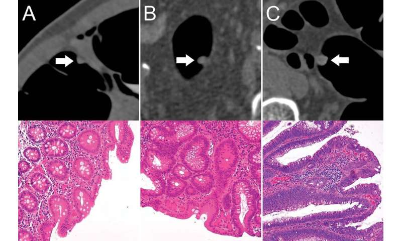
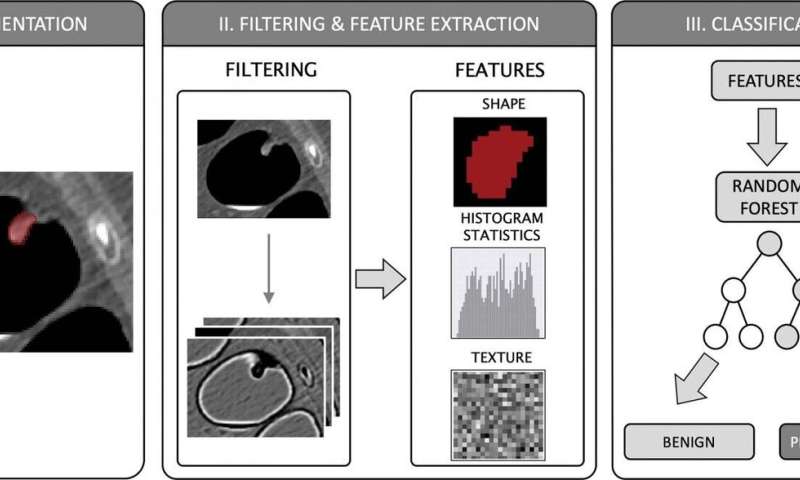
 in the descending colon of a 78-year-old woman. B, Virtual fly-through three-dimensional reconstructions were used for exact polyp localization. C, Manual polyp segmentation was performed in multiplanar two-dimensional CT colonography images. D, CT colonography images were preprocessed for image feature extraction by application of a dedicated filter. Credit: Radiological Society of North America")
A machine learning algorithm helps accurately differentiate benign and premalignant colorectal polyps on CT colonography scans, according to a study published in the journal Radiology.
Colorectal cancer is among the three most common causes of cancer-related death among men and women in industrialized countries. Most types of colorectal cancer originate from adenomatous polyps—gland-like growths on the mucous membrane lining the large intestine—that develop over several years. Early detection and removal of these precancerous polyps can reduce the incidence and mortality of colorectal cancer.
During the last two decades, CT colonography emerged as a noninvasive alternative to colonoscopy in screening for colorectal cancer. It is comparable to colonoscopy in detecting most polyps and is effective at visualizing portions of the colon that in cases of complex anatomical conditions cannot always be evaluated by colonoscopy. However, CT colonography does not enable a definite differentiation between benign and premalignant polyps, which is crucial for individual risk stratification and therapy guidance.
For the new study, researchers leveraged the power of radiomics, a process of extracting quantitative features from medical images, to characterize polyps beyond what was apparent to the naked eye.
The researchers developed a machine learning algorithm to predict the character of the individual polyps based on quantitative image features extracted through radiomics. They applied the noninvasive, radiomics-based machine learning method on CT colonography images from a group of asymptomatic patients at average risk of colorectal cancer. The machine learning algorithm was trained on a set of more than 100 colorectal polyps in 63 patients and then tested on a set of 77 polyps in 59 patients.
In the test set, the machine learning approach enabled noninvasive differentiation of benign and premalignant CT colonography-detected colorectal polyps, with a sensitivity of 82%, and specificity of 85%. The area under the curve (AUC), a graphical measurement that reflects how much the model is capable of distinguishing between benign and precancerous polyps, was excellent.
“These results serve as proof-of-concept that machine learning-based image analysis allows the noninvasive differentiation of benign and premalignant colorectal polyps in CT colonography data sets,” said study lead author Sergio Grosu, M.D., radiologist from University Hospital, Ludwig Maximilian University of Munich, in Munich, Germany. “The AUC of 0.91 indicates that this method works well.”
The findings point to a role for machine learning-derived algorithms in boosting the effectiveness of CT colonography as a screening tool for colorectal cancer.
“Adding machine learning-assisted image analysis to conventional, radiological image reading could further improve the clinical significance of CT colonography-based colorectal cancer screening by allowing for a more precise selection of patients eligible for subsequent polypectomy,” Dr. Grosu said. “This method could be used routinely as a second reader in all CT colonography examinations in the distant future.”
Dr. Grosu said that additional studies with larger numbers of patients are needed to validate the findings. He added that these studies should also help drive improvements in the machine learning algorithm.
Source: Read Full Article