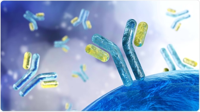
A research group, funded by CSL Behring and led by Marc Bürzle, have published a paper in which they describe a fluorescence-activated cell sorting (FACS)-based method for isoagglutinin quantification.

Image Credit: Utas7777777/Shutterstock.com
This FACS method offers a more precise means of quantifying immunoglobulins for intravenous application and confirms differences in intravenous immunoglobulin products.
Isoagglutinins molecules found in intravenous immunoglobulin (IVIG) products are linked to a process called hemolysis, the rupturing of red blood cells. Isoagglutinins are antibodies that stimulate the ‘sticking together’ or agglutination of red blood cells and are typically anti-blood group A and B antibodies.
As such, knowledge of the number of isoagglutinin is necessary to control their levels and determine their correlation with hemolytic events.
The previous quantification of isoagglutinin has been outlined by the European Pharmacopoeia (Ph.Eur.) in a direct assay and has since been considered the standard for assessing isoagglutinin levels in IVIG products. In this assay, IVIG samples are subject to papain-treated A1 or B blood group red blood cells (RBCs), after which the level of agglutination is visualized.
Agglutination is measured by a cell-pattern which is produced upon two-fold dilutions of IVIG products mixed with a +/- one titer step. The results are variable, with actual concentrations between half as much as the test result and double the amount. Consequently, the correlation between the isoagglutinin level and hemolysis is difficult to establish.
The Study
The group, headed by Marc Bürzle, has developed the FACS-based method which can quantify the isoagglutinin level in the IVIG level with higher precision than the current Ph/Eur. direct assay. This FACS-based approach expands on a methodology outlined by Gerber et al, with the addition of an external reference standard and a bioassay.
This includes evaluating several concentrations of a series of standard and test samples for comparison of similarity between them – the similarity is a crucial assumption for bioassay analysis. Bioassays are commonly used in the release of drug products and characterizing intermediates and formulas.
The group report results from a range of IVIG and subcutaneous immunoglobulin (SCIG) products as determined by the assessment of isoagglutinin levels. This method has also overhauled the process of the IVIG production process. Initially, donations from donors with high anti-A titers were excluded; a more efficient immunoaffinity chromatography (IAC) step replaces this.
IAC reduces anti-A and anti-B antibodies with minimal impact on antibody specificities in the final product without causing loss of the total IgG concentration.
Method
To achieve this, red blood cells (RBCs), erythrocytes, positive for the antigens A1 and B and negative for RH ccddee were used from three donors from the Interregional Blutspende SRK AG, Bern, Switzerland.
Phenotypes of the A1 and B RBCs were verified using a test to check for agglutination using anti-A and anti-B antibodies respectively. The FACS assay was performed by a flow control cytometer, and from each IVIG sample (either standard or control), a set of 3 concentrations were produced as a result of two independent two-fold serial dilutions.
Each of the testing solutions was incubated for an hour on a microtiter plate, and following incubation, each of the microtiter plates was centrifuged and once again subject to incubation. Following this second round of incubation Hey PBS buffer was added, and the median fluorescence intensity of each test solution was measured via flow cytometry.
Results
The results demonstrated start the quantification of isoagglutinin IGIV products could be determined would high precision. Relative to the Ph. Eur direct assay, this new method shows less variability – this is because of the endpoints on the older assay measured on a discrete scale, at low resolution. Because this assay is based on FACS, the results are continuous (as well as electronic and objective) and can compensate for the effect with Ph. Eur.
Owing to the objective readout of the FACS assay. the results are automated and processed electronically, circumventing the objective interpretation needed in the Ph. Eur. assay. The team's new essay was also able to differentiate between different isoagglutinin levels in the commercially available IVIG products tested, as well as in products before and after being subject to isoagglutinin reduction strategies.
The assay was sensitive enough to be able to detect variability present in the same batch. Most importantly, Bürzle et al.’s analysis was able to demonstrate that there are potentially clinically important differences between IVIG products, however, due to a lack a representative sample testing, conclusions about isoagglutinin quantities could not be formed.
In addition to this, when the team performed screening and exclusion of samples or using the IAC agglutination reduction step, the anti- A levels in the IVIG product were reduced.
The use of the IAC reduction step brought down the levels to those similarly seen using the old method of Cohn-like ethanol fractionation, in which the majority of isoagglutinin are segregated into a fraction.
At present, the levels of anti-A and anti B must be equal to or lower than the control – the result of the groups IVIG testing shows that someone concentrations in products are higher than 100% of the standard.
The acceptability of the release of these products is due to the variability using the old Ph. Eur direct assay. The group suggests that by implementing the new assay, the release of batches with higher isoagglutinin levels would be prevented, hence increasing the stringency of the specification.
Bürzle and colleagues recognized that their method was limited in that the isoagglutinin content of the standard material was not known. Also, there is no international reference preparation present for anti-A and anti-B quantification.
As a result, isoagglutinin concentrations can only be expressed relative to a standard. Further, the test it's not 100% specific for each of the antibodies, and the team cannot exclude the presence of non-ABO antibodies binding to other RBC antigens – these are called irregular antibodies.
The team excluded the contribution of irregular antibodies based on the fact that there was a strong correlation between the Ph. Eur direct assay and FACS-based method, however.
Future Research
While previous research on the correlation between isoagglutinin levels in IVIG products and the risk of Hemolysis is lacking, the FACS based approach offers the opportunity to improve this. This is because previous methods could not measure with precision.
In total, this new FACS-based assay allows for the quantification of isoagglutinin both in products manufactured using chromatography-based and Cohn-fractionation- based products, in addition to the efficiency of isoagglutinin reduction as a result of IAC.
The high precision afforded by this method is hoped to result in more stringent isoagglutinin specifications for products, as well as being able to investigate the correlation between Hemolysis and isoagglutinin content in IVIG products.
Funding
This work was funded by CSL Behring.
Declaration of competing for interest
All authors are currently, or were previously, employed by CSL Behring, the funders of this research. Also, Alphonse Hubsch and Dominik Stadler own shares of CSL. There are no further conflicts of interest to report.
Acknowledgments
Editorial support was provided by Meridian HealthComms Ltd, funded by CSL Behring.
Source
Bürzle, M. et al. Measurement of isoagglutinin in immunoglobulins for intravenous application by flow cytometry. Analytical Biochemistry. 2019. DOI: https://doi.org/10.1016/j.ab.2019.113534
Further Reading
- All Flow Cytometry Content
- Flow Cytometry Methodology, Uses, and Data Analysis
- Flow Cytometry Techniques used in Medicine and Research
- Flow Cytometry History
- Fluorescence-Activated Cell Sorting
Last Updated: Jan 31, 2020

Written by
Hidaya Aliouche
Hidaya is a science communications enthusiast who has recently graduated and is embarking on a career in the science and medical copywriting. She has a B.Sc. in Biochemistry from The University of Manchester. She is passionate about writing and is particularly interested in microbiology, immunology, and biochemistry.
Source: Read Full Article