
History
Causes and symptoms
Epidemiology
Case reports Diagnosis and treatment
References
Further reading
Chanarin-Dorfman syndrome (CDS) is a rare, multisystemic, neutral lipid storage disease (NLSD) where the triglyceride accumulates in the leukocytes and other organs.

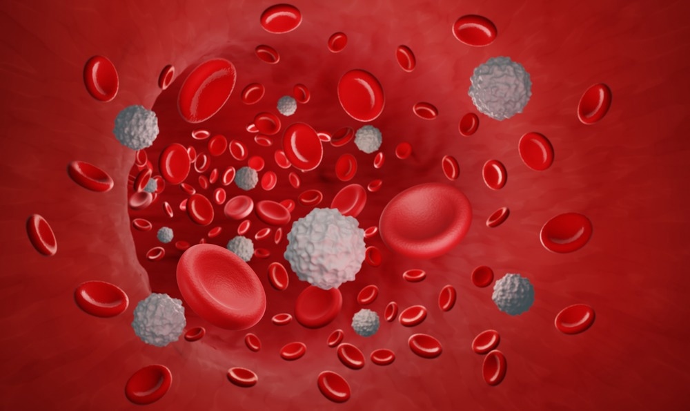
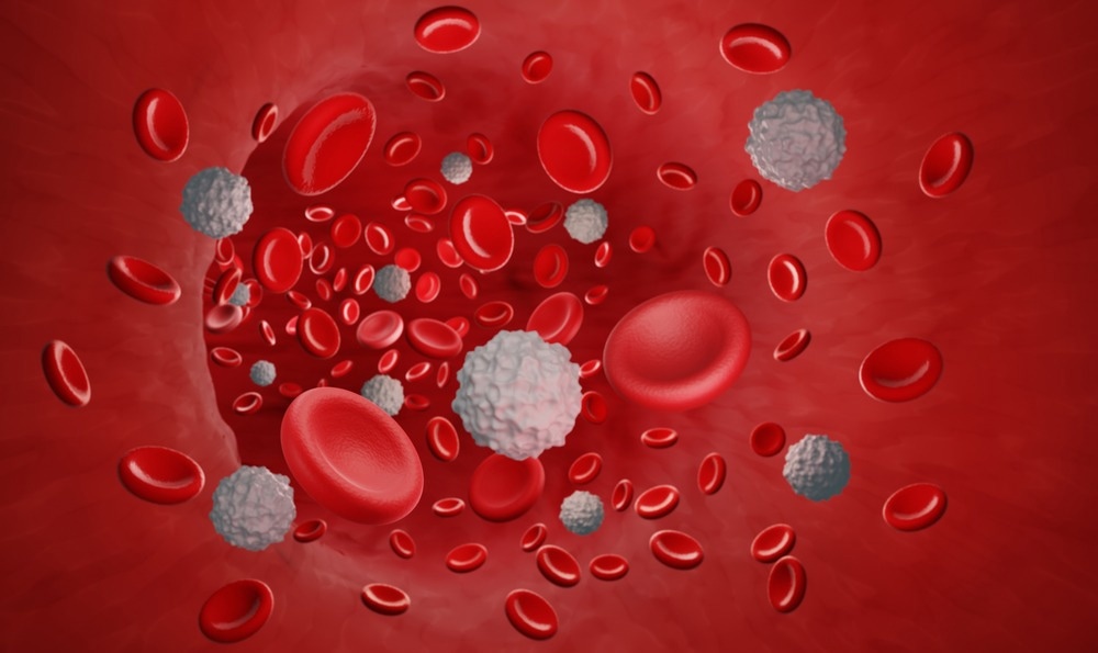
Image Credit: ART-ur/Shutterstock.com
First described by Dorfman in 1974, the syndrome follows an autosomal recessive inheritance pattern. It is caused by a mutation in the ABHD5 gene and is associated with the deposition of cytoplasmic neutral lipid droplets at multiple sites, including the liver.
History
Jordan discovered lipid vacuoles in the cytoplasm of leukocytes from the peripheral blood smear of two brothers with progressive muscular dystrophy for the first time in 1953. Dorfman and Chanarin, two decades later (in 1974 and 1975, respectively), reported for the first time a neutral lipid storage disorder defined by lipid accumulation in peripheral blood leukocytes, bone marrow granulocyte precursors, liver cells, and a variety of other cells in the body.
Causes and symptoms
The abhydrolase domain containing the 5 (ABHD5) gene, also known as the comparative gene identification-58 (CGI-58) gene, is located on the 3p21 chromosome and causes the syndrome. This gene produces a protein of 349 amino acids and a molecular mass of around 39 kD that activates adipose triglyceride lipase (ATGL), which plays a crucial role in intracellular lipolysis.
The ATGL enzyme is completely or partially inactivated when the ABHD5 gene is mutated. This enzyme allows the release of fatty acids (FAs) from the intracellular triacylglycerol (TG) reserves of adipose and non-adipose tissues. This results in insufficient FA mobilization within the cell, resulting in the formation of TG-containing intracytoplasmic lipid droplets.
Non-bullous congenital ichthyosiform erythroderma and hepatic steatosis are the most common clinical symptoms of CDS. Other symptoms include splenomegaly, sensorineural hearing loss, nystagmus, ataxia, myopathy, cataract, mental retardation, and growth retardation. These clinical symptoms are likely to vary depending on the patient's ethnic origin and the type of mutations.
Muscle involvement is common (about 60% of cases), and symptoms include slowly progressing weakening of proximal limb muscles while axial muscles remain unaffected, elevated serum muscle enzymes, and electromyographic evidence of myopathy.
Epidemiology
Around 120 cases of CDS have been recorded around the world to date. According to a study, the prevalence of CDS is similar in men and women, albeit men are somewhat more likely to develop it. In terms of age, the youngest patient was six months old, while the oldest was 68 years old. Nearly half of persons with this disease have consanguineous parents worldwide.
Furthermore, the rate of consanguineous marriages is higher than the global average in countries/regions such as Turkey, India, and the Middle East. As a result, the proportion of homozygotic and heterozygotic patients in these areas is higher than in other areas.
Case reports
Waheed et al., in 2016, described a one-year-old boy who had skin lesions and hepatomegaly since birth. He was the result of a consanguineous union. He met all of his developmental milestones and was breastfed until he was six months old. His cognitive, neurological, and ophthalmological exams were all normal. Steatohepatitis was discovered in a liver biopsy. Jordan's abnormality, a persistent hallmark of Chanarin-Dorfman syndrome, was validated by a peripheral blood smear analysis.
Demir et al., in 2016 described a 15-month-old girl with CDS who exhibited ichthyosis, elevated liver enzymes, hepatomegaly, liver fibrosis, and an ABHD5 mutation. She complained of dry skin and itching. Her parents were second-degree relatives. On physical examination, erythema and fine scaling were noted on the patient's skin. She also experienced abdominal distension and liver hypertrophy.
Hepatomegaly was discovered on abdominal ultrasonography. Histopathological examination indicated lipid accumulation in the parenchyma of the liver, hydropic degeneration in the remaining parenchymal cells, and a significant increase in fibrous tissue in the portal area. Triglyceride levels were found to be elevated in laboratory tests.
Hawat et al., in 2021, presented the first documented case of splenectomy in a CDS patient. Congenital ichthyosis was diagnosed at birth, and the child was treated locally. When he was seven, he developed hazy vision in both eyes and was diagnosed with Splenomegaly. They performed a splenectomy surgery owing to pancytopenia, and after three months of monitoring, his hemoglobin, WBCs, and RBCs all improved significantly. This case suggests that when CDS patients develop hypersplenism and pancytopenia, splenectomy may be a good treatment option.
Diagnosis and treatment
The presence of ichthyosis and the discovery of lipid droplets in granulocytes (Jordan's abnormality) in a peripheral blood smear are used to diagnose. Sequencing the ABDH5 gene in patients diagnosed with CDS can help confirm the diagnosis.
Patients with this syndrome do not have a specific treatment. A diet high in carbohydrates, medium-chain fatty acids, and ursodeoxycholic acid (UDCA) is recommended for CDS patients. An oral retinoid, such as acitretin, may be given to patients with severe ichthyosis. In these patients, topical administration of urea-containing emollients is also effective. UDCA works against cholestasis through a variety of methods, including anti-inflammatory, immunomodulatory, and antiapoptotic.
The patients were given a 5-year low-fat, enriched, medium-chain fatty acid diet, which was proven effective in treating skin disorders and restoring the liver to its normal size. For patients with CDS and decompensated cirrhosis, liver transplantation may be an option. However, such patients will still require diet therapy and long-term monitoring.
References
- Cakmak, E., & Bagci, G. (2021). Chanarin-Dorfman Syndrome: A comprehensive review. Liver international: official journal of the International Association for the Study of the Liver, 41(5), 905–914. https://doi.org/10.1111/liv.14794
- Hawat, M., Khouri, L., & Almansour, M. (2021). Splenectomy in Chanrain-Dorfman syndrome. Journal of Pediatric Surgery Case Reports, 68, 101822.
- Kalyon, S., Gökden, Y., Demirel, N., Erden, B., & Türkyılmaz, A. (2019). Chanarin-Dorfman syndrome. The Turkish journal of gastroenterology: the official journal of Turkish Society of Gastroenterology, 30(1), 105–108. https://doi.org/10.5152/tjg.2018.18014
- Demir, B., Sen, A., Bilik, L., Deveci, U., Ozercan, I. H., Cicek, D., & Dogan, Y. (2017). Chanarin-Dorfman syndrome. Clinical and experimental dermatology, 42(6), 699–701. https://doi.org/10.1111/ced.13163
- Waheed, N., Cheema, H. A., Suleman, H., Mushtaq, I., & Fayyaz, Z. (2016). Chanarin-Dorfman Syndrome. Journal of the College of Physicians and Surgeons–Pakistan: JCPSP, 26(9), 787–789.
- Nur, B. G., Gencpinar, P., Yuzbasıoglu, A., Emre, S. D., & Mihci, E. (2015). Chanarin-Dorfman syndrome: Genotype-Phenotype Correlation. European journal of medical genetics, 58(4), 238–242. https://doi.org/10.1016/j.ejmg.2015.01.011
- Bruno, C., Bertini, E., Di Rocco, M., Cassandrini, D., Ruffa, G., De Toni, T., Seri, M., Spada, M., Li Volti, G., D'Amico, A., Trucco, F., Arca, M., Casali, C., Angelini, C., Dimauro, S., & Minetti, C. (2008). Clinical and genetic characterization of Chanarin-Dorfman syndrome. Biochemical and biophysical research communications, 369(4), 1125–1128. https://doi.org/10.1016/j.bbrc.2008.03.010
Further Reading
- All Rare Disease Content
- What is a Rare Disease?
- Importance of Research into Rare Disease
- Teaching old drugs new tricks – drug repurposing for rare diseases
- What is Adenomyosis?
Last Updated: Sep 4, 2023